Blog Layout

A1 Medical Imaging to Be Part of Artificial Intelligence Software Development for Medical Diagnostic Imaging Industry

A1 Medical Imaging to Be Part of Artificial Intelligence Software Development for Medical Diagnostic Imaging Industry 


From the desk of Peter Solodko, CEO

A1 Medical Imaging 


SARASOTA, FLORIDA – May 20, 2021 – Marilyn Radakovic, RN, Chief Operating Officer at A1 Medical Imaging, is excited to announce the company will become an integral part in the development of Artificial Intelligence (AI) software products for the Medical Diagnostic Imaging Industry. This program, which is just getting started, is being spearheaded by Consolidated Healthcare Services (CHS), a company that is led by its CEO, Peter Solodko. CHS provides administrative management services to all A1 Medical Imaging clinics.


Artificial Intelligence has the potential to extrapolate meaningful information from raw data. Part of this CHS project involves extracting relevant information from large amounts of data so that predictive modeling can be developed. A1 Medical Imaging has years of accumulated imaging data that can facilitate and expedite this development process.


The goal will be the development of a suite of AI software products that will enhance the identification of abnormalities within patients. The suite will include products to improve imaging outcomes, report findings and back-office systems.


“A1 Medical Imaging has been in business since 2008 and has multiple imaging centers in Florida and Georgia,” Marilyn Radakovic said. “We have an enormous amount of imaging data to be scrutinized.”


“Utilizing A1 Medical Imaging’s data is exactly what’s needed to make this venture viable,” Peter Solodko added. “If not for that, the success of this project would be severely challenged.”  


The program is just getting underway, beginning with the identification of its key contributors including CHS managing the project, skilled software developers that are highly knowledgeable about Artificial Intelligence, and A1 Medical Imaging with its large volume of data.


Solodko went on to say, “Our management group has been providing administrative services to the medical diagnostic imaging industry since 2004. We have the essential knowledge to manage this project. By combining with the programming skills of the software developers, and the mountain of data from A1 Medical Imaging, we have all the necessary ingredients to make this vision a reality.”


To learn more about A1 Medical Imaging, visit www.a1mri.com.


 About A1 Medical Imaging

 A1 Medical Imaging has become a model of excellence in the diagnostic industry. From its home office in downtown Sarasota, Florida, the company maintains primary in-house functions of administration, human resources, transcription, finance, insurance contracting, information systems and technology, billing and collections, and medical technology. Utilizing “state of the art” computer technology, A1 Medical Imaging can monitor the overall performance and operation of each center from its one central location, successfully assuring cost-effectiveness and efficiency throughout the company.


 Media Contact: 

 Ronald G Hock


A1 Medical Imaging LLC


Email: rhock@chsmed.com

By 7073618507 09 Jan, 2023
MRI scans are performed to look at the internal structures of the body. The two types of MRIs are open MRI and closed MRI. The traditional MRI involves the patient lying on a table inside a giant tube. The radio waves and giant magnets in an MRI create an image of the internal structures of the body from every angle. Musculoskeletal MRI Musculoskeletal MRI is a common diagnostic test used for evaluation of musculoskeletal conditions. The techniques used to create MR images have advanced over the years. Commonly used pulse sequences have also been enhanced. To properly interpret these images and to improve future imaging protocols, it is important to understand the physics involved.  Musculoskeletal MRI uses radio waves and a powerful magnetic field to produce detailed images of your body. These images are extremely sensitive and allow your doctor to accurately diagnose a variety of conditions. These images can show inflammation and bone erosion. In addition, they can help your physician determine the cause of your pain. Musculoskeletal MRI is often part of the diagnostic algorithm for most muscle injuries. Because muscle MRI images can be easily confused with normal anatomical structures or pathological signs, interpretation of these images must be done by a multidisciplinary team. This article aims to reduce common misinterpretations of these medical images. Before you have a Musculoskeletal MRI scan, it is important to remember that this is a procedure that requires the patient to lie still for long periods of time. Young children may require sedation to make them more comfortable during the scan. Fortunately, the process is painless and requires no blood or other invasive procedures. Musculoskeletal MRI scans are generally performed as an outpatient procedure. The technician will position you in a narrow circular tube in order to capture images of the bones and soft tissues. This scan can be done at home or in the hospital. It can take 15 to 45 minutes to complete. The results of this test will be reviewed by a radiologist, who will interpret the results and notify your primary care physician. Musculoskeletal radiology is a branch of radiology that deals with the imaging of bones, cartilage, connective tissue, and muscles. Its experts have special training and expertise in the diagnosis and treatment of diseases affecting the joints, muscles, and spine. They are considered leaders in their field and have a strong track record of patient outcomes. Musculoskeletal MRI scans can be very helpful in diagnosing various disorders. Work-related disorders and sports-related injuries are a few common examples. Other disorders may include tumors in the soft tissues surrounding the joints and extremities. In addition to cancer, a Musculoskeletal MRI scan can be used to evaluate the health of the spinal cord following a trauma. A Musculoskeletal MRI scan involves the use of a powerful magnetic field and radio frequency energy to produce detailed pictures of the internal organs and tissues. The procedure is painless and noninvasive. The results of the scan are used to plan treatment and monitor its effectiveness. The scan process usually lasts between 15 and 45 minutes, depending on the body part being examined. The differences between a Musculoskeletal MRI scan and a CT scan are often quite similar. The differences between the two are due to the differences between water and fat protons. Therefore, understanding the differences between the two types of MRIs can help you better distinguish between a symptomatic lesion and a nonsymptomatic one. Functional MRI Functional MRI scans are a powerful diagnostic tool for analyzing brain functions. These scans can reveal brain structures that appear normal but are faulty, and can also reveal areas in the brain that have undergone significant changes. This type of imaging is used in hundreds of scientific articles each month, and is also often referenced in the lay press. Functional MRI scans measure brain activity by combining multiple images taken less than a second apart. The scan works by measuring changes in blood flow within the brain during specific tasks. The results are useful for brain mapping and assessing the risk of injury and brain surgery. They can also be used to determine whether certain diseases or conditions are causing changes in the brain. Functional MRI scans have a better spatial resolution than MEG and EEG, which are both low-resolution methods. The spatial resolution of a typical MRI is typically millimeters, but there are ultra-high-resolution scans, which can reach tens of micrometers. These MRI scans use 7-T fields and fine iron oxide to ensure the highest possible resolution. Functional MRI scans are highly complex, requiring a lot of statistical analysis. At first, this complexity caused many to mistrust the results of functional MRI scans. However, as researchers became more familiar with the technology, these studies have become more reliable. A functional MRI scan can give a doctor a good idea of where to operate on the brain to restore function. The MRI scanner produces a strong magnetic field, and it is recommended to avoid wearing metal items that could get in the way of the imaging process. Moreover, the area of the body that will be imaged will be slightly warm. If you experience any discomfort, it is best to inform the radiologist as soon as possible. For the best results, the patient should be perfectly still while the images are being recorded. The entire procedure typically takes a few minutes. A functional MRI scan is an imaging test that measures brain activity by using blood oxygen level-dependent contrast. During the test, the blood in the brain is oxygenated and deoxygenated, which creates differences in the magnetic susceptibility of these two types of blood. Functional MRI scans are commonly performed as a part of brain activity research. In the most common fMRI scans, different slices of the brain are acquired at different times. This allows researchers to compare the signals recorded during different states. The results of the analysis are used to create an activation map of the brain. In contrast to fMRI scans, functional MRI scans are noninvasive and do not involve any surgery or injections. Rather, they are a valuable part of brain mapping research. Open MRI Open MRI scans are similar to closed MRIs but are more comfortable for patients. Patients who are claustrophobic or who are sensitive to loud noises may find them uncomfortable. However, newer machines are getting closer to producing images of the same quality as closed MRIs. Patients still need to lie still, and the open MRI scan type does not completely enclose the patient. Open MRI machines are more accommodating to larger patients with claustrophobia. These MRI machines have front and back windows and are usually open on two sides. They are also more advanced than closed MRI machines, but take longer to produce images. Patients should consider the size and comfort of the MRI machine before choosing it. Open MRIs are not as useful for detecting small or delicate body parts. But they are useful for detecting lesions in the brain and wrist, and can provide doctors with a more detailed diagnosis. They can also be used to detect single or multiple vessels in patients with coronary artery disease. While an MRI scanner does not use radiation, the magnetic field is intense. The electromagnetic waves that travel through the body cause the atoms to temporarily move. The atoms emit different amounts of energy, which vary depending on the type of tissue. The MRI scanner captures the energy generated by the hydrogen atoms and converts it into a picture. The images can help diagnose any problems in a person's body. Open MRI scans are quieter than closed MRIs and are also better for larger patients. Open MRIs are also more child-friendly, and a parent can be present when a child is having an MRI. They are also safer than closed MRIs. Open MRI scans are commonly used for a variety of purposes. These types of MRIs are very useful for identifying symptoms of stroke, heart diseases, disc disease, bone infections, and more. They are also useful for diagnosing internal organs. And unlike traditional MRIs, they are able to produce more detailed images in less time. Open MRI scans can be performed on people with claustrophobia, and the procedure is more comfortable for the patient. As the MRI machine is a circle, the patient can move into the circle's center opening, making it 20% larger than conventional MRI scanners. Additionally, most patients are able to lie on their feet while receiving the exam, making it more comfortable for them. MRI scans highlight contrasts in soft tissues, and this makes it easier for physicians to identify problems in joints, cartilage, tendons, and ligaments. They also help identify infections and inflammatory problems. They can also help physicians rule out tumors. These scans are also useful for assessing the anatomy of the brain. Open MRI scans can be performed in a variety of positions, and are the most common MRI scan types. There are a number of pulse sequences available for these types of imaging.
By 7073618507 09 Jan, 2023
If you're wondering if you need a CAT scan, you've come to the right place. This article will explain what a CAT scan is, how it works, and what to expect during the procedure. You'll also learn about the different types of CAT scans available, including those with contrast and those without. CAT scans CAT scans are performed to get a clearer picture of your body. A contrast medium is given to help make images more visible. This can be given as a drink or through a small tube into your arm. It will stay in your arm until the scan is complete. Your health care provider can recommend a facility that offers these procedures. You can also check with the American College of Radiology to see whether the radiologist you choose is accredited.  CT scans use a computer to create a multi-slice image of the body. They can reveal structural problems and abnormalities in nearly any part of the body. The most common types are CT scans of the chest, abdomen, and head. These scans take a few minutes and require little or no anesthesia. They can also detect lung cancer. The scans are painless and noninvasive, but do have a small risk of radiation. A CT scan of the chest can reveal a patient's lungs in great detail. If a patient complains of chest pain or difficulty breathing, a doctor may order this scan to determine the condition of the lungs. A CT scan can help diagnose various lung conditions, including aortic aneurysm, emphysema, and lung cancer. CAT scans with contrast CAT scans with contrast are a type of diagnostic test that makes body tissues easier to see. The contrast agent is a liquid that is given to the patient through a thin tube into the arm. It stays in the body until the scan is complete. Patients may also take an oral sedative called Buscopan prior to the scan to ease muscle spasms. While contrast material is usually harmless, some people are allergic to it and experience mild itchiness or rash. In extreme cases, a reaction can be life-threatening. Your health care provider will monitor you after the CT scan to ensure that you do not have a reaction. You should also tell your doctor if you are taking any medication that may cause allergic reactions. A CAT scan is one of the most accurate diagnostic tools available. It allows doctors to examine the head, neck, chest, pelvis, and abdomen. Different types of tissues exhibit different degrees of natural contrast, which improves the quality of the image. However, soft tissues that are in contact with blood are harder to image. For these reasons, contrast imaging is often used to improve the visualization of soft tissues. Some CAT scans require contrast material, which helps highlight soft tissue on the scan. Contrast material is not a dye, but instead works by blocking the X-rays and appearing white on the image. However, it is important to note that some people may be allergic to the contrast material. Contrast is usually administered via an intravenous (IV) line. This line will be inserted into a vein in the lower spine or arm. The IV line will be left in the patient until the scan is completed. The contrast agent may cause a brief flushing or a metallic taste. Some contrast materials may cause complications in the kidney. If you have kidney problems, you should notify your doctor before having a CT scan. The dye may leak into the bloodstream and affect the kidneys. In this case, you should take extra fluids and medicines to help your kidneys eliminate the dye safely. It is also important to watch your diet during the four hours before the CAT scan with contrast. Drinking clear liquids such as black tea, broth, and ginger ale is important to prevent nausea caused by the contrast dye. Aside from liquids, patients should avoid taking oral diabetes medications 48 hours before the scan.
By 7073618507 09 Jan, 2023
The benefits of ultrasound are well known. They include being noninvasive, reducing the need for invasive procedures, and cost. They also have no unpleasant side effects. Hence, many physicians prefer this method to traditional procedures. Whether you're a newbie or a seasoned physician, ultrasounds are a great option for assessing your health. Non-invasive The use of non-invasive ultrasound technology can be helpful for many reasons. Non-invasive methods avoid the risk of surgery, radiation, and postoperative pain. They also are cheaper and easier to use. Unlike MRIs, non-invasive ultrasound does not require an incision or injection.  Ultrasounds are useful tools for doctors, because they can image internal organs. While X-rays are best for imaging bones and other hard tissues, ultrasounds can image the lining of the lungs and other soft tissue. They are also able to image blood vessels and the lining surrounding the heart and other organs. Furthermore, they are useful for detecting conditions such as liver fibrosis, which results from inflammation. Therapeutic ultrasound uses sound waves above the human hearing range to target, heat, or break up diseased tissue. HIFU (High Intensity Focused Ultrasound) is another method of using ultrasound to target and destroy diseased tissues. This technology is not invasive and, because of this, it is safer for both the mother and the unborn child. A non-invasive ultrasound may also be less painful for the patient. The entire procedure lasts about 15 to 30 minutes. The results may be available immediately or a few days later. During the ultrasound, there are no side effects, but some people experience some discomfort afterward. A few minutes of discomfort is normal but should go away after the scan. The ultrasound technician will apply water-soluble gel to the affected area of the body. This gel does not stain the patient's clothing or harm the skin. A handheld transducer moves over the gel and creates images of inside the body. The patient will be asked to remain still for several seconds and may be asked to hold their breath. After the ultrasound, the technician will remove the gel from the skin. Another non-invasive ultrasound uses Doppler technology to determine blood flow patterns in the body. This technology can detect any blood flow problems by detecting the direction and speed of the blood flow in the body. This type of ultrasound can also detect blood flow problems in the carotid arteries, as well as the blood flow to the brain. Non-ionizing radiation Ultrasound treatment is considered a non-ionizing radiation. Unlike ionizing radiation, which has the potential to cause cancer, non-ionizing radiation does not alter the atoms and molecules of the body. This means that there is no risk of DNA mutation or cancer. However, non-ionizing radiation can interfere with electrical devices in the body, such as an artificial heart pacemaker. It can also damage tissue. The use of non-ionizing radiation is regulated by the UNIRSIG, a global body that ensures that medical practitioners follow current regulations and practices. This includes the use of ultrasound and the use of MRI. Other non-ionizing radiation applications include fiber optics and magnetic resonance scanning. Studies have shown that ultrasound can have a range of benefits, including detecting congenital malformations during pregnancy. However, more studies are needed to determine the risks of ultrasound treatment during pregnancy and in the womb, and the effects of ultrasound and contrast media on infants. Ultrasound imaging is a non-ionizing radiation imaging modality that provides high-resolution images of the internal body. The technique is used to diagnose conditions such as tumors and blood vessels in the pelvic region. It can also be used for image-guided interventions on the musculoskeletal system. Ultrasound imaging uses non-ionizing radiation because it does not involve ionizing particles. Ultrasound imaging uses optical fibres and advanced lens systems that are non-ionizing. Unlike X-rays, non-ionizing ultrasound imaging does not affect internal organs. While ultrasound is not the only non-ionizing radiation treatment available to physicians, it has been shown to be very effective. It is commonly used for diagnosis. During pregnancy, ultrasound can also be used to detect abnormalities and placental location. Low-intensity Doppler ultrasound is also used to monitor fetal heart rate. Ultrasound technology has been widely adopted in the last 50 years. Today, almost all pregnancies involve some exposure to ultrasound. Although most research focuses on the higher exposure levels, there are some adverse effects associated with ultrasound treatment. Ultrasound is a high-frequency sound wave that can penetrate the body and provide information about the structure and functioning of organs. Its wavelength allows it to selectively absorb energy from different body tissues. In the future, it may be used to destroy cancer cells. Convenience Ultrasound scans are used to assess the health of your fetus during pregnancy. They're painless and can provide quick results. A trained technologist applies a gel to the skin and moves a transducer over the area of your body that needs to be examined. When the sound waves bounce off body structures, they create an image that is then viewed on a monitor. These images can then be printed or stored electronically. Since ultrasound uses sound waves, there is no risk of long-term exposure, so this procedure can be used on women and small children alike. Ultrasound scans can help determine the cause of many different medical problems. Most healthcare providers recommend having an ultrasound at least once during a pregnancy. These images are useful for tracking a fetus's development throughout the nine months. They may also reveal the biological sex of the fetus. An ultrasound study is usually performed by a certified radiologist. The technologist will apply a water-soluble gel to the area of the body that needs to be studied. The transducer is a small handheld device that sends sound waves through the body to produce images. Afterwards, the radiologist will review the images and send them to the doctor who ordered the test. Typically, an ultrasound test takes about 30 minutes to an hour. While ultrasound is not dangerous, there are some risks. Exposure to ultrasound energy is not advised for non-medical purposes, and untrained users are likely to pose a risk of radiation damage. It is important to follow the physician's instructions before undergoing an ultrasound. They will also inform you of any necessary preparations. Some ultrasound apparatuses display the image in three dimensions. This improves the convenience of ultrasound diagnosis. The ultrasound apparatus can automatically select a region of interest (ROI) if a user selects a point within the tumor. The ROI is displayed in a three-dimensional format that allows a physician to analyze it accurately. Cost The cost of ultrasounds can vary widely depending on the location. For example, if you live in a small city and don't have health insurance, it may cost more to go to a hospital than to have an ultrasound done in a larger city. However, if you have health insurance, the costs of an ultrasound can be covered by your health insurance plan. If you don't have health insurance, you'll need to contact your health insurance provider to find out if you're covered. If your insurance doesn't cover ultrasounds, you'll have to pay for the entire procedure yourself. Alternatively, you can negotiate the cost of your ultrasound directly with the provider. In some cases, an ultrasound is covered by health insurance, but you'll still need to pay the deductible and coinsurance. In addition to location, the cost of an ultrasound will also depend on the provider you choose. There are many different types of ultrasounds. Some are diagnostic and others are therapeutic. Diagnostic ultrasounds are usually more expensive than therapeutic ultrasounds. Hospitals also tend to charge higher prices than private outpatient facilities. A diagnostic ultrasound involves high-frequency sound waves being emitted into the body and reflected back to the ultrasound transducer. The reflected sound waves are captured by the transducer, which then makes the image visible. When you buy an ultrasound machine, you should also consider the additional costs associated with training and installation. Many ultrasound suppliers include basic installation and training in their purchase prices. However, if you want detailed training and instruction, you may want to hire a specialist. This training can run anywhere from $1,000 to $6000. Another thing to keep in mind is that ultrasound machines require many different types of consumables, such as transmission gel, pads, and lotions. The cost of an ultrasound can vary depending on where you live and where you get it. In some areas, an ultrasound can cost over $1,000. You should check with your health insurance provider to see if your coverage covers the procedure.
By 7073618507 14 Nov, 2022
During a PET scan, the body's cells emit radiation called positrons, which are radioactive elements that can be used to detect disease. This method is noninvasive, painless, and enables physicians to detect disease at an early stage. It also helps detect areas of activity inside the body. Noninvasive A Noninvasive Positron Emission Tomography (PET) scan uses a small amount of radioactive tracer to visualize the blood flow and function of heart muscle tissue. The tracer is injected into a vein in the arm and travels to the heart where it is detected by special cameras. The process produces images of the heart at rest and when the patient is given medication. It helps doctors diagnose certain heart conditions and certain types of cancer.  A PET scan is noninvasive and generally painless. The procedure involves a small dose of radioactive material that is attached to a common compound. This is usually glucose, though other compounds similar to glucose can also be used. The patient receives the tracer through a vein and a specially designed positron scanner uses the images to obtain detailed information about the body's metabolism of the tracer. PET has been in clinical use since the early 1990s. The most common tracers used in a PET scan are rubidium-82 (Rb) and nitrogen-13-ammonia (N-ammonia). This procedure takes less time than a SPECT scan, and the radiotracers used in a PET scan have shorter half-lives than the SPECT scan. The test can be completed in less than 30 minutes. The main goal of a PET scan is to diagnose the cellular changes that occur in the body during disease. The PET scan uses a small amount of radioactive material to create images. The radioactive tracer is similar to sugar but is attached to a small radioactive atom. The radioactive substance travels through the bloodstream to the patient's brain and is detected by brain cells as positrons. A PET scan is an excellent diagnostic tool. It can show whether a patient is suffering from cancer and whether it has spread throughout the body. It is also an important part of a patient's care, and can help doctors plan treatment options early. Painless A Painless Positron Emission Tomography (PET) scan is a type of medical test in which a radioactive substance is injected into the body through a small plastic tube. The patient lies still on the scan table for 30 to 60 minutes. The radioactive substance, known as FDG, travels through the blood to the area of the body being studied. The substance is then absorbed by the tissues, and the scan can take place. This scan provides doctors with a detailed view of the human body and organs. It helps determine organ function by measuring the metabolism of organs and tissues. It also allows physicians to see how cancerous cells affect various organs. It is also an effective tool for detecting brain tumors and heart disease. A PET scan uses radioactive substances to create images of the body's organs and systems. It is most commonly used to detect cancer, monitor the progression of the disease, and determine the effectiveness of chemotherapy. But it can also be used to diagnose a variety of other diseases, including cardiovascular diseases and brain disorders. PET scans are relatively painless. They involve injecting a tracer liquid into a vein, usually in the arm. The patient may also swallow a tracer or breathe it in. The liquid then travels throughout the body and collects in an organ. As it moves through the body, it gives off positive-charged particles (positrons). A camera records the positrons, turning them into pictures on a computer screen. PET tests can help doctors assess the effects of trauma on the brain, assess the level of perfusion in the heart muscle, and assess the spread of heart disease. Unlike other types of tests, a PET scan does not require anaesthesia or sedation. Patients need to be completely still for the procedure to be performed. PET scans are generally outpatient procedures. This means that a patient doesn't have to stay overnight in the hospital. However, patients are encouraged to follow up with their doctor if they are ill before their appointment. Can detect disease in its earliest stages Using a special camera and a substance called a "tracer," a positron emission tomography scan can detect cellular changes in the body. This procedure is used to diagnose heart, lung, and brain conditions. Compared to other imaging tests, the images from a PET scan can detect disease in its earliest stages. The process of positron emission tomography involves injecting a small radioactive material into the patient's body. This radioactive substance helps the doctor create a 3-D picture of the organs in a person's body. These scans can detect the earliest stages of many types of disease and help doctors determine the effectiveness of treatment. The scans can also reveal abnormalities in organs and parts of the body, including tumors and brain disorders. The technology behind positron emission tomography is based on the physical properties of isotopes. They emit positrons during decay. A PET center uses special radiochemical laboratories to produce these isotopes. These radioactive substances travel through the bloodstream and enter the patient's brain. Once inside, they bind with ligands in brain cells and then release positrons into the body. In a PET scan, a positron emitter is passed through a detector pair. These detectors detect coincidence events in the positrons emitted. They are made from bismuth germinate or lutetium oxide and contain photomultiplier tubes. Signals from these detectors are then fed into separate amplifiers and energy discriminating circuits. This technique is able to pinpoint disease in its earliest stages. In addition to the earliest detection of disease, PET scans can also help in the treatment of a variety of diseases, including cancer. PET scanners can detect disease at its early stages by measuring the metabolism of cancer cells. The FDG-F-18 positron emitter is an important workhorse in tumor imaging. Tumor cells increase their expression of glucose transporter molecules and hexokinase in their tissues, which makes it possible for them to consume more FDG. Can detect radioactive glucose A Positron emission tomography (PET) scan can detect cancer by detecting the levels of radioactive glucose in the body. It works by using a small amount of radioactive glucose, known as a tracer, to produce computerized images of the body's chemical structures. PET scans can detect cancer in several ways, including tumor detection, and they can be used in conjunction with a CT scan to enhance the accuracy of detecting abnormalities. A PET scan uses a radioactive substance called FDG, or fluorodeoxyglucose, to detect tumours and other medical conditions. The substance accumulates throughout the body and gives off gamma rays that are detected by the PET scanner. Once the scan is complete, patients slide out of the machine. They can normally return to their daily activities, though they should limit their activity for 30 to 90 minutes after the scan to reduce the risk of radiation exposure. Patients should avoid eating and drinking for several hours before the scan to avoid affecting their sugar metabolism. Patients should also refrain from strenuous exercise 24 hours before the appointment. The appointment letter will include instructions on how to prepare. The radioactive tracer is only active for a brief time, so patients should avoid eating or exercising for at least 6 hours before the scan. Patients should wear loose-fitting clothing and take off any metal jewelry before the scan. The PET scan takes 30 to 60 minutes. After the patient is ready, he or she will be given a contrast infusion through an IV or CVC. This medication is then slowly infused into the patient's bloodstream. The patient must remain silent and still for 30 to 60 minutes before the scan. Afterwards, the doctor will look over the results. The patient can be told within a week of the scan.
By 7073618507 07 Nov, 2022
A CT scan is an imaging exam used to detect internal organs and other structures that are not visible to the naked eye. It can help diagnose diseases such as cancer, heart disease, and emphysema. It can also help identify liver masses. However, discussing the CT scan with your doctor before you undergo it is important. Cancer There are many benefits to a CT scan, and they can even help detect cancer. But what are the risks associated with a CT scan? One of the biggest risks is radiation. A CT scan produces more radiation than X-rays, so it is best to avoid the procedure if you are pregnant. But you must carefully weigh these risks against the benefits of a CT scan.  The advantage of a CT scan is that it provides more detailed information than X-rays. This is especially true when contrast is used. The contrast makes certain tissues more visible. Cancer cells absorb the contrast, making them appear white on the scan. This allows the radiologist to better interpret the images. The contrast also helps him, or her see organs and tissues surrounding a lesion. There are also risks involved with CT scans for cancer. Children are particularly susceptible to radiation. However, they typically live longer than adults. Children should only undergo CT scans when they have a clear clinical indication. Heart disease A CT scan is an important screening test for heart disease. It can detect the presence of plaques in the coronary arteries. Whether these plaques are solid or soft, CT scans can help physicians determine whether patients have coronary artery disease. The scan results can be used to make informed decisions about treatment. The procedure takes only a few minutes, and patients can resume their daily activities afterward. A CT scan can help doctors determine the location of blockages in the heart arteries, which is important for preventing a heart attack. However, this test does not replace traditional risk factors assessments, which can help identify patients at risk for cardiovascular disease. Therefore, doctors should ensure that the radiation risks do not outweigh the benefits of a CT scan before recommending it to patients. A CT scan is an imaging test that uses multiple rotating X-ray beams that are guided by a computer to produce a clear image of the heart. Patients are asked to wear loose-fitting clothing and remove any metal objects that could affect the images. Additionally, they may be asked to stop eating or drinking before the test. In addition, certain medications should be stopped before undergoing a cardiac CT scan. Emphysema The use of computed tomography (CT) scans in diagnosing and monitoring emphysema has increased tremendously since the mid-1980s. Emphysema is a condition of permanent enlargement of airspaces and destruction of alveolar walls in the lung. While pathologic examination of tissue is limited to post-mortem specimens, CT scans provide non-invasive structural information on the lung. Today, CT scanners are readily available in virtually every hospital in the developed world. The technique is particularly useful for emphysema diagnosis, as it can identify lung volumes that are not normal. An emphysema CT scan can detect this abnormality if the lung's attenuation value is less than -960 HU. The amount of kilovoltage, collimation, and reconstruction algorithm are important parameters in the acquisition of CT scans. CT scans provide high-quality images of the lungs, which is valuable for clinical and research purposes. The images provide a clearer picture of the lung's structure and can help distinguish between emphysema and other lung diseases. Liver mass Performing a CT scan to identify a liver mass can reveal various information. The contrast agent, Mn-DPDP, can help doctors better detect abnormalities. However, contrast-enhanced CT can miss a malignant hepatic lesion in some cases. One example of a CT scan that missed a malignant hepatic lesion was in a patient with a single 2.8-cm metastasis to the liver. CT scans are more detailed than standard X-rays. For instance, they can provide more information about liver cancer. They also allow doctors to determine whether cancer has spread elsewhere. In addition, CT scans can be used to evaluate the biliary tract. A liver and biliary tract CT scan can also help doctors perform biopsies. A CT scan may also detect incidental findings that may not appear on a physical exam. This can be a sign of a tumor that is not cancerous or an enlarged liver. A recent study of over 17,000 individuals found that 6 percent of patients had incidental hepatic findings. The incidental findings were typically benign, although some patients had multiple lesions. Bleeding The benefits of CT scans in bleeding patients are numerous. The scans help determine the exact location of the bleeding and may aid in diagnosing or excluding certain conditions. They can also identify enteric tract pathology, define postoperative bowel anatomy, and evaluate vascular disease. Before undergoing a CT scan, patients should consult with their doctor about any current medications. Also, they should tell their care team about any allergies that they have to minimize the risk of a reaction. A CT scan may require a contrast dye, which is injected to highlight specific areas of the body. This contrast agent may be injected intravenously or administered orally through an enema. CT scans can also help detect GI bleeding. Unlike traditional x-rays, CT scans send a series of small x-ray beams through the body. They combine these beams to form a cross-sectional image. These images are important in diagnosing various diseases and can often eliminate the need for exploratory surgery. Pain CT scans are a fast and painless way to diagnose various illnesses and conditions. A radiologist with specialized training can detect a range of conditions with high accuracy and may reduce the need for more invasive procedures. CT scans are particularly effective in detecting internal injuries and bleeding. They also provide detailed images of various types of tissue. Patients are generally asked to lie still for a CT scan. They may be asked to shift positions during the test to obtain different body views. Typically, CT scans do not cause pain, although you should alert your technician if you experience any pain during the test. The test will be stopped if you experience discomfort. Non-invasive CT scans are fast and painless, offering detailed images of the body's bones, soft tissues, and blood vessels. These scans are particularly useful in diagnosing internal injuries and bleeding. Additionally, they are a cost-effective imaging method, often costing less than MRI. Another benefit of CT scans is that they allow for the reconstruction of images in an infinite plane, giving doctors a more detailed view of the body. Before undergoing a CT scan, you must let your physician know about any medications or medical conditions you may have. For example, if you have an allergy to a specific medication, tell your healthcare team about it. In addition, if you have a history of severe or life-threatening allergic reactions to medications or contrast agents, tell your care team about them so that the procedure can be conducted safely and effectively. Another benefit of CT scans is that they can reveal structures of nearly every body part, including organs, blood vessels, and other tissues. This means that they can help physicians diagnose diseases and even guide minimally-invasive procedures. For example, a CT scan can identify tumors or abnormalities in the abdomen, search for tumors in the urinary tract, and diagnose cardiac conditions, including aneurysms and heart disease. Fast A fast CT scan uses advanced computer technology to visualize the entire body. It can detect blockages and abnormalities leading to a heart attack or chest pain. It displays unique slices of the body, making it a more accurate diagnostic tool than an X-ray. It has the added benefit of being extremely detailed, allowing a doctor to see if a condition is serious enough to warrant further testing. A CT scan can also help doctors diagnose injuries to the head and spine, as well as liver and lung diseases. It can also detect blood clots and internal bleeding. It is most often used when a rapid diagnosis is essential, such as during an auto accident. The scan can even save a life in an emergency department. A CT scan can help doctors make faster diagnoses and improve workflow in the emergency room. By reducing the time between an emergency department patient arriving and undergoing a CT scan, doctors can identify and treat conditions with more effective therapies.
By 7073618507 31 Oct, 2022
An X-Ray is a diagnostic imaging test that helps a doctor identify many different health conditions. They can reveal fractures and infections, as well as help monitor your condition. These tests can also be used for a number of other purposes. In this article, you'll discover some of the main benefits of an X-Ray. X-rays are a diagnostic imaging test X-rays are diagnostic imaging tests that use small amounts of radiation to obtain images of internal structures. The exposure from X-rays is safe for most adults, but it is not recommended for pregnant women. Women should tell their doctor if they are pregnant and ask if they can use ultrasound instead. X-rays can be used to detect broken bones and other problems with the body.  They can also help detect cancer and other life-threatening conditions. Depending on the type of x-ray used, patients may be exposed to varying levels of radiation. It is important for pregnant women to discuss this with their health care providers before getting an X-ray because the radiation can harm the fetus. Patients generally do not feel any pain during X-rays, but the technician may ask you to hold your breath for a few seconds while the image is taken. Children may have difficulty staying still, so their provider may use a restraint to keep them still during the procedure. These restraints can reduce the need for multiple retakes and are safe for your child. X-rays are a painless diagnostic imaging test that doctors use to get a clear picture of internal structures. The X-rays are produced through the body's tissue and clothing, and a digital image is generated. The images are a series of negative and positive images that show internal structures. In the case of broken bones, a doctor can use this to determine the exact location of the fracture. Other applications include the assessment of bone fractures and tumors. X-rays are an excellent tool in diagnosing many ailments. They can be extremely helpful in revealing the location of tumors or other abnormalities. While most patients have heard of x-rays as a way to look for fractures, a doctor can use this test to check the structure of a bone. X-rays can be taken at home or in a hospital. A radiologist or X-ray technologist will position the patient to obtain an accurate image. They may use pillows or sandbags to hold the patient still during the imaging process. They also use a contrast dye, which helps in highlighting specific areas of the body on the X-ray images. They can reveal fractures The X-Ray image is an important tool for physicians to use for fracture diagnosis. X-Rays can help identify bone fractures and prevent further damage. Moreover, X-rays can help determine fracture severity. X-ray images are often required to diagnose fractures in children. X-Ray images are produced by exposing the affected area to radio waves. The radiation emitted by x-rays is very small. This means that the risk of getting cancer from X-rays is negligible. The procedure allows physicians to see fractures, tumors, masses, enlargements of certain organs, and foreign objects. However, this procedure has risks. X-rays are not only used to diagnose fractures, but can also help detect infections, dental decay, and even bone cancer. They can also detect foreign objects that have become lodged in soft tissue. X-Rays are quick and easy to use, and they are useful in emergency situations. Besides, the procedure is very safe and does not cause any side effects. However, patients should avoid wearing metal jewelry while undergoing the procedure. The X-rays can also reveal abnormalities in the joints. Your doctor will analyze the images to determine if you have fractures or foreign objects. The images are also useful in assessing bone growth in children. Besides, x-rays can reveal fractures and abnormalities in the joints. X-rays are also useful during certain procedures, such as coronary angioplasty. These X-rays help doctors guide a catheter through arteries, allowing them to do their work more effectively. Besides, they do not require any preparation on your part. You can eat and drink normally before the procedure and continue to take your usual medicines. Nevertheless, some patients are advised to refrain from certain drugs before their X-rays. They can reveal infections X-Rays can reveal infections in various parts of the body. Some infections can remain undetected for weeks or even months, so it's important to get the correct diagnosis early. These images can also reveal any changes in a bone or joint. In addition to revealing infection, x-rays can reveal fractures. They can help monitor medical conditions X-rays are an important imaging tool that can diagnose and monitor medical conditions. While some scientists have expressed concern over the health implications of X-rays, they do offer many benefits. For instance, they can monitor conditions like fractures and tumors that are causing discomfort or pain. The use of X-Rays is not prohibited by law but it is important to know how much radiation you're exposed to. X-Ray imaging devices must comply with federal and state regulations. The FDA's guidelines and regulations apply to all X-ray imaging facilities. In addition to helping doctors diagnose medical conditions, X-Rays can also monitor and track the progression of treatment. The images from these tests can also show unusual features or pathology, which can help doctors plan the proper course of treatment. Moreover, an X-ray can detect certain types of cancer. While the chances of developing cancer from X-rays are not great, they are still high compared to the natural incidence. Although X-rays are often used to detect broken bones, they can also be used to visualize organs in the chest and lungs. X-rays are made using a specialized machine that produces a digital image. Digital images are processed faster than traditional X-rays, reducing the risk of error. Moreover, these images are stored electronically. An X-Ray procedure can take as little as 15 minutes, but it can take longer depending on the complexity of the problem. During the process, the X-ray images are recorded on a computer. The radiologist will interpret the images and send the results to your doctor. Your physician will explain the results to you and schedule follow-up visits. If you're unsure about the process, you should ask questions. X-Rays are an excellent diagnostic tool that allow doctors to see the internal structure of the body without the need for surgery or other treatments. They provide a clear image of internal structures and can detect fractures, tumors, and even cancer. Moreover, they are inexpensive and can help physicians monitor various medical conditions. Computed tomography, also known as CT, is an imaging method that combines computer processing and x-ray technology to create three-dimensional images of the body. CT images are much more detailed than plain x-rays, allowing doctors to see structures from multiple angles.
By 7073618507 24 Oct, 2022
An MRI scan is a safe, painless test that can show the inside of the body. It helps doctors distinguish different types of tissue because protons in the tissue realign differently and produce different signals. The signals from millions of protons combine to create a detailed picture of the interior of the body. Patients should not feel any discomfort during the test and can even lie down during the procedure. However, those who suffer from claustrophobia may need to ask a radiographer how to deal with their fear of the process. MRI scans are non-invasive An MRI scan is a type of imaging test that uses magnetic resonance imaging (MRI). An MRI scan doesn't involve radiation or surgery, and is therefore a great option for many patients. However, this type of test is not without risks. It can take up to an hour to perform, and the patient must remain still during the entire procedure.  An MRI scan can reveal problems that other tests cannot detect. It can be a valuable tool for the early diagnosis and evaluation of many focal lesions. The technology is extremely sensitive and can detect abnormalities that are hidden by bone. In addition, MRI scans are excellent for assessing the biliary system, as they can reveal abnormalities in this area that aren't visible on other types of imaging. Although an MRI scan is very safe, it is important to note that there are some potential risks. The FDA receives about 300 reports of adverse events per year, and most of these describe problems related to heating or thermal injury. The most common problem reported by patients is second-degree burns, but other issues include projectile events and falls. It is also important to note that some patients experience nerve stimulation during the MRI. MRI scans are not dangerous, but they can be stressful for people who have an extreme fear of enclosed spaces. However, sedation and other medications can be used to keep patients calm during the scan. Patients should follow the instructions of the facility to minimize potential side effects. MRI scans are also useful for tracking treatment. They can reveal changes in organ, lesion or tumor size. In addition, they can reveal abnormal blood flow in organs. This information can help doctors diagnose suspected tumors and determine how best to treat them. An MRI scan can reveal differences in water content and blood flow between different tissues. A cancerous tumor may have an abundance of new blood vessels that supply the tumor with more blood than the surrounding tissue. They are painless An MRI is a safe and painless procedure. Patients are usually given a gown and asked to remove any jewelry or glasses. The exam may take a few minutes to complete. Before the test, patients may be given a sedative or anesthesia that will help them relax. A nurse will be present to help them. After the exam, patients will be taken to the recovery room. While an MRI is a painless procedure, you will need to lie still during the procedure. If you move during the scan, the images will be blurred. The scan itself will last about 15 minutes. Some patients may experience claustrophobia, so earplugs or headphones can be helpful. Patients should inform their physician if they are pregnant or have any metal implants before getting an MRI. Although an MRI does not cause any pain, the procedure is noisy, so it is important to wear headphones or earplugs. Patients should also remove all magnetic objects from their body before going into the machine. The procedure itself will last between half an hour. After the procedure, the patient can resume normal activities. However, people who are afraid of being in a closed space can ask their healthcare provider about medication that will help them relax. A mild sedative will help you sleep, but it is necessary to follow all instructions closely. During the procedure, patients will lie on a table that slides inside an MRI machine. The table may be equipped with a plastic coil that wraps around the head. The technician will then take several pictures of the brain. Each picture takes a few minutes. The patient will be able to communicate with the technician through a microphone. They are safe for people who are vulnerable to radiation While there are some concerns that MRIs are not safe for people who are vulnerable to radiation, some studies have shown that they are safe. This study involved a nationwide survey in England where 85 patients with leukemia and 45 unaffected controls were compared. The results showed that the difference between the two groups could not have been fortuitous. However, the study also indicated that radiation from MRIs may initiate changes in a fetus or young child. The risk of cancer from imaging-based radiation is small compared to the overall risk of cancer. It is estimated that the risk of cancer from a single CT examination is approximately 42 per 100 patients. These small risks must be multiplied by a large population to find the overall risk. Even if the patient's dose is not excessively high, the risk of radiation-induced death is not high, averaging about 50 per 100,000 CT patients. In 2003, the FDA classified MRIs up to 8 T as nonsignificant risk devices for nonneonatal patients. During the same period, the International Commission on Non-Ionizing Radiation Protection determined that acute exposures at this level caused no serious health effects. In addition, the International Electrotechnical Commission increased the static magnetic field limit of the first-level controlled operating mode to eight T. One vendor even received a CE mark for a 7-T clinical system. People who are sensitive to radiation should be aware that MRIs are safe for those with genetic conditions. Because MRIs are painless, claustrophobia or other medical conditions shouldn't interfere with the scan. They can show both bones and soft tissues MRI, or magnetic resonance imaging, is a diagnostic test that creates cross-sectional images of your body. These images are created by a computer and magnetic field. They are a good way for doctors to view both soft and hard tissues in the body. MRIs are more detailed than x-rays and can show diseased tissues and tumors. The radiation used during x-rays is absorbed by dense matter, such as bone, but passes through less dense soft tissues. In a typical x-ray, several angles are taken and the images are compared to those of the uninjured limb. The entire process usually takes about 10 minutes. The images are then developed and written to a CD, or can be viewed on a computer screen. MRIs are also useful in diagnosing bone injuries. They provide detailed images of the bones and soft tissues in the body, such as the cartilage. The scan also helps doctors identify bone spurs, which are often difficult to detect with X-rays alone. An MRI can also show inflammation of soft tissues, which are not possible to detect through X-ray images. Before an MRI, patients must be completely still and remove any jewelry or credit cards. Patients should also bring any relevant X-rays and an insurance identification card. An MRI can be difficult for people who are claustrophobic, but an open MRI can help you relax. MRIs use contrast media to improve the visibility of soft tissues and improve diagnosis. The contrast agent is not the same as the dye you might use at home. MRIs use non-iodine gadolinium-based contrast agents. They're safe and effective. But some people may be sensitive to these drugs and a kidney function test may be required. In addition, metallic substances in the body can affect MRI images and cause discomfort or injury. Therefore, it's a good idea to consult with your physician about any medical history before scheduling an MRI. They are faster than X-rays An MRI is a diagnostic imaging procedure that uses radio waves and a magnetic field to create detailed pictures of internal organs, bones, and tissues. While X-rays are more commonly used to detect broken bones, an MRI can also detect diseased tissues, such as tumors. Furthermore, the radio waves used in MRIs are far safer than x-rays because they carry much lower levels of energy than visible light rays. Another major difference between an X-ray and an MRI is the speed of scanning. An MRI can take only a few seconds, while an X-ray can take minutes. And because MRIs are faster, they are also more sensitive. This means that they're often more useful in diagnosing cancer, especially in young children. Moreover, MRIs are safer and cheaper for adults, too. The MRI is also faster than a CT scan. CTs use ionizing radiation to create a 3D image of a region, and they are also less expensive than X-rays. X-rays are better for certain types of conditions, such as fractures and serious injuries, while MRIs are better for diagnosing internal organs, such as kidney disease. X-rays are faster than MRIs, but they're not without their own disadvantages. Patients must lie motionless for an MRI to give the best images. This is because movement may cause blurry images. Depending on the type of scan, MRIs can take as little as 15 minutes or as long as an hour. The speed of MRIs has increased dramatically in the last decade. The fastest brain scans can now take less than a half second. The newest MRI technology combines x-rays with a computer. This allows researchers to get a three-dimensional picture of the brain in less than a half second, compared to two to three seconds for the typical X-ray. This speed is critical for neuroscience.
By 7073618507 17 Oct, 2022
Medical imaging uses a variety of techniques. These include X-rays, Computed tomography, MRI, sonography, and more. You can find out more about these types of tests below. These types of tests help doctors diagnose certain conditions by producing detailed images that a standard X-ray cannot. X-rays X-rays are used in medical imaging for a variety of purposes. The radiation produced by an X-ray machine passes through most solid objects in the body to produce an image. This image is then used to help healthcare providers visualize internal structures of the body. Some radiographs include contrast medium, which is given to patients intravenously, orally, or rectally. These contrast mediums are usually colored white or grey, depending on their density. Bone and other solid objects appear white, while skin, muscle, blood, and fat are gray.  Although X-rays are a common imaging modality, the risk of exposure to ionizing radiation remains low. There are a number of laws in place to ensure the safe use of X-rays. These laws are implemented by various government regulatory agencies and advisory bodies. Understanding these laws may help radiologic technologists make sound decisions regarding the use of x-rays in medical imaging. Although X-rays are safe for most people, they may increase the risk of developing cancer if repeated exposures are made over the course of a lifetime. Therefore, it is important to discuss your risk with your healthcare provider before scheduling an appointment for an x-ray. Additionally, it is important to remember that pregnant women should never hold their babies during an x-ray. A number of x-ray imaging methods are now available. Single-frame x-ray tomosynthesis (SRS) captures up to 30 images per second and provides a 10 to 100 times higher spatial resolution than conventional tomosynthesis. This method could lead to a safer, less invasive way to treat lung tumors and cardiovascular diseases. X-rays are electromagnetic radiation with a very short wavelength and high energy. As such, they can penetrate through bones and soft tissue. In addition, they don't carry a charge. Unlike visible light, x-rays are not affected by magnetic fields, so they do not interfere with magnetic field measurements. They are also capable of producing a photoelectric effect, which makes them a popular tool for medical imaging. Today, medical imaging is the largest application for X-rays. It is used for image-guided therapy and medical diagnostics. The technology behind X-rays is based on vacuum electronics and a mechanism known as bremsstrahlung. It was first discovered more than 120 years ago, and since then, it has become a valuable medical imaging tool. MRI MRI medical imaging is a common medical procedure, but there are certain precautions patients should take before having the procedure. For instance, pregnant women should let their MRI technologist know if they are pregnant, and should leave jewelry and metal at home. They should also bring a photo ID and insurance card. The MRI machine is a tunnel-like device that has a powerful electromagnet. This magnet forces the hydrogen atoms in the patient's body to align with its magnetic field. High-frequency radio waves are then emitted from the magnet, which causes protons to spin, and then attempt to realign with the magnetic field. This motion causes an electric signal, which is captured by a sensor. If you're pregnant, it's best to let your MRI technologist know before you have your exam, because some of the contrast material is toxic to unborn children. It's important to disclose this to the technologist so that he can advise you on how to safely receive the contrast agent. MRI medical imaging is often used to diagnose and treat a variety of conditions. Different MRI sequences may be useful for different purposes, such as assessing the cerebral cortex, identifying fatty tissue, and characterizing focal liver lesions. Some MRI sequences can be performed simultaneously using these techniques. MRIs can be used to diagnose conditions such as herniated spinal discs, multiple sclerosis, and liver cirrhosis. These images are acquired in a special digital format called DICOM. DICOM ensures high-quality images. In addition to the quality of the images, MRIs can provide highly accurate data. Before undergoing an MRI medical imaging procedure, patients must remove all metal objects from their bodies. They should also wear a cotton gown. During the procedure, patients are required to remain still. A radiography technologist monitors the process from a separate room. Patients can talk to the technologist via a microphone, which helps ease the fear of being confined in a narrow tube. The procedure can take anywhere from 15 minutes to an hour. MRI medical imaging is typically painless. The duration of the exam varies depending on the type of study that a patient needs. The actual test time is between 20 and 60 minutes, though prep time can extend the appointment time. The time may also be increased if multiple studies are needed. Sonography A sonogram is a form of medical imaging that uses ultrasound waves to visualize internal structures. The sonographer uses a transducer that moves over the patient's skin and then captures images. The images are then displayed on a computer screen. The sonographer will review the images with the radiologist, who will interpret the results and provide the doctor with a report. The patient will be asked to remove any clothing that may be obscuring the area being examined. Then, the sonographer will ask the patient to lie on a bed. There are many types of sonography, including vascular and neurosonology. Vascular sonographers create images of blood vessels and arteries, while obstetric and gynecologic sonographers focus on the female reproductive system. Other specialized sonographers specialize in imaging the abdominal and musculoskeletal systems, as well as the heart. Cardiovascular sonography focuses on cardiac arteries and valves. The benefits of sonography are many. For instance, it can help doctors diagnose pelvic masses, infertility problems, and inflammatory diseases. It is considered one of the most reliable imaging methods. It helps doctors make accurate diagnoses without the use of radiation, surgery, or dyes. Plus, sonography is safe, portable, and non-invasive. Sonography uses sound waves to visualize soft tissue and bone surfaces. It can also be used to diagnose muscle strains and ligament sprains. It can also be used in conjunction with x-ray imaging to detect fractures. Moreover, it can be used to guide a needle during injections. A sonographer must be trained in the use of sophisticated computer equipment. They must also be skilled in working with patients. They must be knowledgeable about anatomy, ethics, and how to communicate with patients. They also have to develop rapport with patients and display confidence throughout the procedure. During the imaging process, a sonographer must have a strong sense of confidence and competence. Another benefit of sonography is the real-time images it produces. This technology is much safer than X-rays, because it does not expose the patient to ionizing radiation.
By 7073618507 05 Oct, 2022
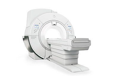
If you have ever had an MRI, then you know that it can be an uncomfortable experience. The equipment in traditional MRIs is shaped like a cylinder, and the patient is positioned inside for the duration of the scan while the technologist speaks with them through a microphone. Although the traditional MRI is the best type of MRI for producing high-quality images, it can be too claustrophobic for some patients.  Young children or those with high BMI should have a parent present during the procedure to protect them. But the True Open MRI allows the patient to move around freely, allowing for a more comfortable experience. Additionally, these new techniques produce higher quality MRI images and better health outcomes. Closed One of the most important factors when choosing an MRI machine is your comfort level. Some patients experience claustrophobia while inside the narrow bore of a closed MRI machine. This is especially true for obese or stocky patients who may be uncomfortable in the small confines of the machine. Another consideration is the time required for a full scan, which can take minutes. In addition, there's no room to move inside the MRI machine, which can obstruct images and result in repeat scans. Moreover, closed MRI machines have a small interior dimension, which prevents large patients from fitting into them. Another important factor to consider when choosing an MRI is the strength of the magnetic field. MRIs with high-powered magnet fields produce more detailed images. However, open-bore machines have lower-quality images than closed-bore systems. Open An open MRI system produces lower-resolution scans than a closed MRI. Because the magnets in open systems can't separate fat from water, the images produced by them are less detailed. Furthermore, the open MRI system takes longer to scan a particular area of the body. This makes open MRI scans less useful for smaller body parts. One of the most important considerations for a child's diagnostic procedure is the medical staff's level of compassion and understanding. Children should always be treated with care and respect by the radiology staff. Even though an open MRI is a relatively painless procedure, some parents may be concerned about the experience. A child can be positioned in the MRI room with a parent, which can be a great comfort for the child. Another factor to consider is the patient's physical abilities. Some patients are claustrophobic or need to be in a wheelchair while having an MRI. If your doctor is concerned that a patient may be afraid of claustrophobia, an open MRI may be a better option. Semi-open Open MRI is the type of MRI that does not put you into a tube. It uses magnets to generate images of your body, but unlike closed MRI machines, you are not fully enclosed. This type of MRI is also relatively inexpensive. Patients can be positioned in a standing or sitting position. Expectations about open MRI are based on the type of scan you'd like. While open MRIs are highly accurate and reliable, their images can be limiting when it comes to imaging deep structures. Because of this, it's important to talk to your healthcare provider to find the right scan for you. Semi-open MRI: A Semi-open MRI provides more space for patients to breathe during the scan. It's often a more comfortable option for patients who are claustrophobic. It also allows the parents to remain with their children during the scan, which is beneficial for those who have difficulty sitting still. Functional Open MRI scans are extremely beneficial for people with specific medical conditions. For example, they can image joints in various positions without requiring a patient to be put under general anaesthesia. Furthermore, they can be used to diagnose many types of injuries that can delay recovery. For this reason, they are often the preferred method of MRI for patients with certain types of pain or back problems. Open MRI systems have a number of benefits, including more comfort and less claustrophobic conditions. Open MRIs also allow a patient to move freely within the MRI machine, which is advantageous for a variety of clinical applications. However, open MRI scans don't provide the same level of detail as closed MRIs. Open MRI machines still use magnets for their diagnostic purposes, but they are not enclosed. In addition to being more comfortable for patients, open MRI machines allow for more movement and reduced risk of claustrophobia and panic attacks. Furthermore, open MRIs allow patients of all sizes to undergo MRIs. In some cases, they may be the only alternative to traditional MRI machines for the treatment of certain conditions. Low-resolution Low-resolution Open Mri is a type of MRI that uses a magnetic field that is weaker than that of closed MRI machines. As a result, it takes longer to acquire images. Because the magnetic field is weak, the images are less detailed and may result in misdiagnosis and the wrong treatment. The main advantage of open MRI is the comfort it offers, but the images produced by an open scanner are less detailed for the radiologist to interpret. However, a closed MRI machine has a higher magnetic field and provides much higher resolution and clarity. This type of MRI can also identify pinched nerve fibers in the body, which are difficult to see on an open scanner. Low-resolution Open Mri is the most commonly used type of MRI, but it is not the only type. The open scanners produce inferior images, and they may require repeated exams using a high-field or closed MR system. This can be frustrating for the patient and may result in higher costs. Image quality While open MR is not as claustrophobic as its closed counterpart, the image quality is less than that of its closed counterpart. As such, it is less suitable for scanning sensitive areas like nerves, joints, and other vascular structures. However, it is still a viable option for certain types of MRI procedures. Another key difference between open and closed MRI scanners is the type of magnetic field strength. Open MRIs use 0.2 to 1.2 Tesla fields, while conventional MRIs use 1.5 to 3 Tesla fields. Higher field strengths mean thinner sections, a higher resolution, and a higher-quality image. Moreover, the open MRI system is more flexible, allowing for more patient positioning options. This allows for more patient comfort, which translates into more calm and less uncomfortable scans. In addition, this type of MRI is more cost-effective than its closed counterpart, and can save up to 40% on installation and maintenance costs. The main drawback of open MRI is the lower signal-to-noise ratio. Compared to closed MRI systems, open MRIs have fewer magnets that are larger, allowing a lower signal-to-noise ratio. However, open MRI systems are better for patients who are claustrophobic, or who prefer a more natural, upright environment. Feasibility Feasibility of Open MR imaging is one of the biggest challenges facing MR centers today. Its high upfront cost makes widespread adoption of this advanced imaging difficult. Moreover, the COVID-19 pandemic has already cost more than $16 trillion in the US, and pressure is increasing on medical systems to cut spending. To effectively deploy open MR, it is essential to invest in modern, high-performance MRI scanners, skilled operators, and the necessary maintenance. Open MRI machines offer benefits to patients in various ways. One major advantage is their ease of use. Moreover, patients are able to be less claustrophobic in open MRI machines. Another benefit is that they can accommodate large and overweight patients. Moreover, they are able to obtain clear and high-quality images of internal body structures. Another benefit of MRF is its low cost. While the technology was originally designed for high-end MRI systems, it can be implemented on older machines. By doing this, it can allow for robust advanced imaging on less expensive or less powerful scanners. Furthermore, it can reduce the cost of building scanners, electrical power, and cooling infrastructure. Moreover, MRF does not require arrays of receiver coils. Safety Open MRIs are gaining popularity as a safe and effective alternative to traditional MRI machines. The main advantage of this technology is that it offers patients more space and less noise. This makes them more comfortable during a procedure, which is good for patients who experience claustrophobia. Open MRI machines also allow the technicians to keep eye contact with their patients, making the experience more pleasant. There are numerous risks involved in an open MRI, and these must be understood before a patient is scanned. MRIs are dangerous because the magnetic fields present within the machine create a powerful static field. A ferromagnetic object that is bigger than the machine can become accelerated in this magnetic field and pose a significant risk to the patient. This is why the safety training program for MRIs should include detailed information on the relationship between object size and material composition. Some fatal MRI accidents have occurred due to inadequate training. Open MRI machines come with a weight-bearing feature that allows the technologist to tilt the machine for the patient. This feature is especially useful when diagnosing back pain or spinal injuries. It also promotes equity. A closed MRI machine can make the patient feel isolated, which is why many wheelchair-users choose open machines.
By 7073618507 21 Sep, 2022
Open MRIs are one of the most popular types of medical imaging, and they're also one of the most affordable. They're non-invasive and pain-free, and can provide you with an array of images, including softer body parts. If you have questions about open MRIs, contact your physician in Cape Coral Florida today.
More Posts
Share by: